
When it comes to heart health, early detection and timely diagnosis can make all the difference. The heart is our body’s powerhouse, pumping oxygen-rich blood to every organ, and any disruption in its function can lead to severe health issues. Thankfully, modern diagnostic tools like 2D Echo (Echocardiography) and ECG (Electrocardiogram) help doctors assess heart health effectively. But what exactly are these tests, and why are they important? Let’s break it down in simple terms.
Understanding ECG – The Heart’s Electrical Blueprint
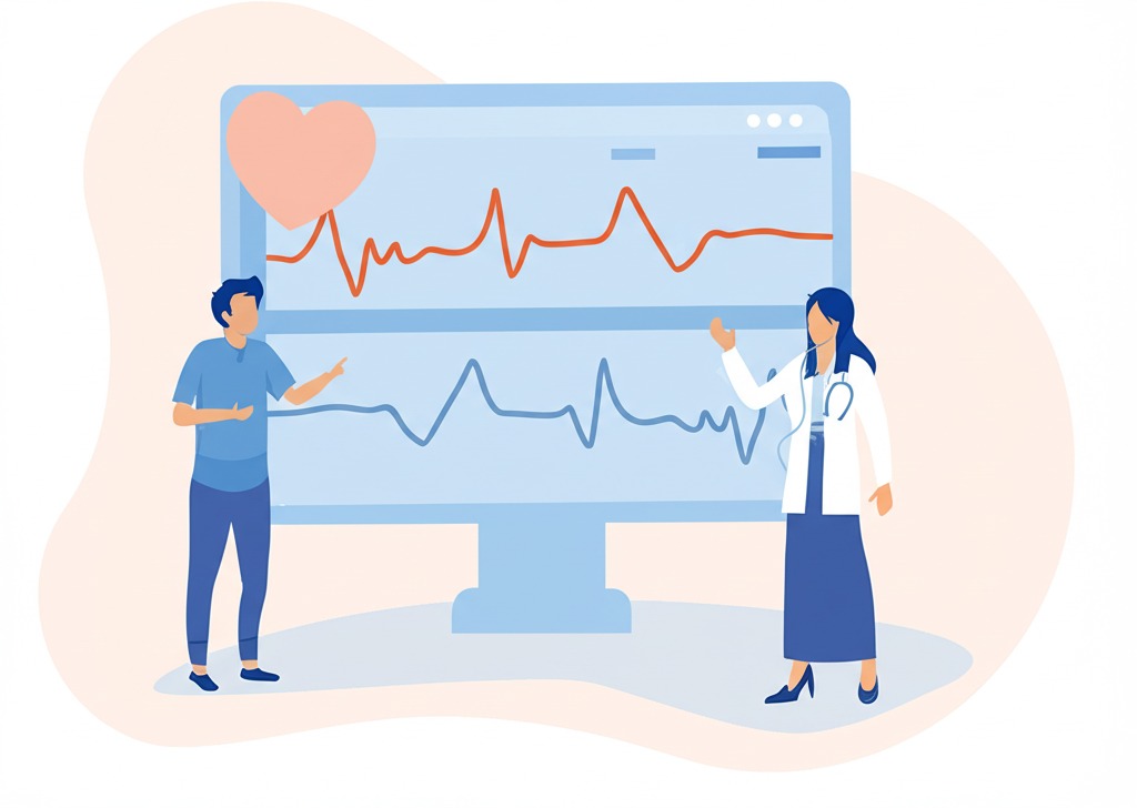
Imagine your heart as a well-synchronized orchestra, where electrical impulses act as the conductor ensuring everything functions in harmony. An Electrocardiogram (ECG or EKG) is a test that records these electrical impulses and helps detect irregularities in heart rhythm.
Why is ECG Done?
ECG is a quick, non-invasive test that helps detect:
- Irregular Heartbeats (Arrhythmias): Conditions like atrial fibrillation, tachycardia, or bradycardia.
- Heart Attack (Myocardial Infarction): ECG can show signs of a past or ongoing heart attack.
- Heart Enlargement: Conditions like left ventricular hypertrophy due to high blood pressure.
- Electrolyte Imbalances: Abnormal potassium or calcium levels affecting heart function.
- Coronary Artery Disease (CAD): Although ECG alone may not confirm CAD, abnormal results can indicate potential heart issues requiring further testing.
What to Expect During an ECG?
- Small electrodes are placed on your chest, arms, and legs.
- The test records electrical activity for a few minutes.
- The process is painless and takes about 5-10 minutes.
Understanding 2D Echo – A Window to Your Heart
If ECG is like a heart’s electrical map, 2D Echocardiography (2D Echo) is like a live video of the heart in action. This test uses ultrasound waves to create real-time images of the heart, showing how it pumps blood and how well the valves and chambers are functioning.
Why is 2D Echo Done?
2D Echo is a crucial tool in diagnosing:
- Heart Valve Problems: Conditions like mitral valve prolapse, aortic stenosis, or regurgitation.
- Heart Failure: Determines how well the heart is pumping blood (Ejection Fraction).
- Congenital Heart Defects: Identifies structural heart issues present since birth.
- Blood Clots and Infections: Detects clots in the heart or infections like endocarditis.
- Pericardial Effusion: Excess fluid around the heart indicating inflammation or infection.
What to Expect During a 2D Echo?
- A gel is applied to the chest, and a transducer (probe) is moved over the heart area.
- The machine captures moving images of the heart in real-time.
- The procedure is painless and takes about 15-30 minutes.
ECG vs. 2D Echo – What’s the Difference?
| Feature | ECG | 2D Echo |
|---|---|---|
| Purpose | Measures electrical activity | Creates images of heart structure |
| Detects | Irregular rhythms, heart attack signs | Valve issues, pumping function |
| Duration | 5-10 minutes | 10-30 minutes |
| Procedure | Electrodes placed on skin | Ultrasound waves used |
| Pain/Discomfort | None | None |
When Should You Get an ECG or 2D Echo?
You may need these tests if you experience symptoms such as:
- Chest Pain or Discomfort
- Shortness of Breath
- Irregular Heartbeats or Palpitations
- Dizziness or Fainting Episodes
- High Blood Pressure or Family History of Heart Disease
- Swelling in Legs (Edema), Suggesting Heart Failure
Regular heart screenings are also recommended if you have risk factors like diabetes, high cholesterol, smoking, obesity, or a sedentary lifestyle.
Take Charge of Your Heart Health
Your heart works 24/7 to keep you going – isn’t it time to take care of it? Whether you need a routine check-up or are experiencing concerning symptoms, Kaizen Diagnostic Centre offers state-of-the-art 2D Echo and ECG services to ensure your heart stays healthy.
📅 Book your heart screening today! Visit www.kaizendiagnostic.com for appointments and expert guidance.


Pingback: Sonography scan What to Expect, How to Choose the Right Lab
Hello! I’m at work surfing around your blog from my new iphone 4!
Just wanted to say I love reading through your blog
and look forward to all your posts! Keep up the excellent
work!
If you wish for to improve your experience just keep visiting this web
page and be updated with the hottest news update posted here.
I would also love to add if you do not currently have an insurance policy or else you do not participate in any group insurance, you could well reap the benefits of seeking the help of a health broker. Self-employed or people with medical conditions usually seek the help of the health insurance brokerage. Thanks for your post.
I got what you intend, regards for putting up.
Pretty nice post. I just stumbled upon your blog and wished to say that I have truly enjoyed browsing your blog posts. After all I will be subscribing to your rss feed and I hope you write again soon!
Thanks for the concepts you have shared here. On top of that, I believe usually there are some factors which will keep your auto insurance premium down. One is, to think about buying vehicles that are in the good report on car insurance providers. Cars which might be expensive tend to be more at risk of being snatched. Aside from that insurance coverage is also based on the value of the car, so the costlier it is, then higher the premium you spend.